Plant problems · hydrangea powdery mildew
Powdery Mildew on Hydrangeas: Identification and Management
Identify powdery mildew on both sides of hydrangea leaves, exclude scorch, spots, and residue, then match cultural management to spread and growth impact.
Published Last updated
In this guide
- Pair both surfaces before naming the disease
- Verify fungal growth across both leaf surfaces
- Map spread and growth impact separately
- Correct crowding without creating sun injury
- Manage vigor from the root zone
- Remove selectively and preserve the canopy
- Keep the treatment claim narrower than the evidence
- Use untreated growth as the next comparison
- Diagnosis
Pair both surfaces before naming the disease
Powdery mildew on hydrangeas produces white to gray surface growth, sometimes with curled or puckered young leaves. On some hydrangeas, yellow, red or purple tissue appears on top while cottony growth is clearer underneath. Inspect paired surfaces across the shrub before choosing management. Crisp margins, bounded lesions and flat residue follow different diagnostic routes.
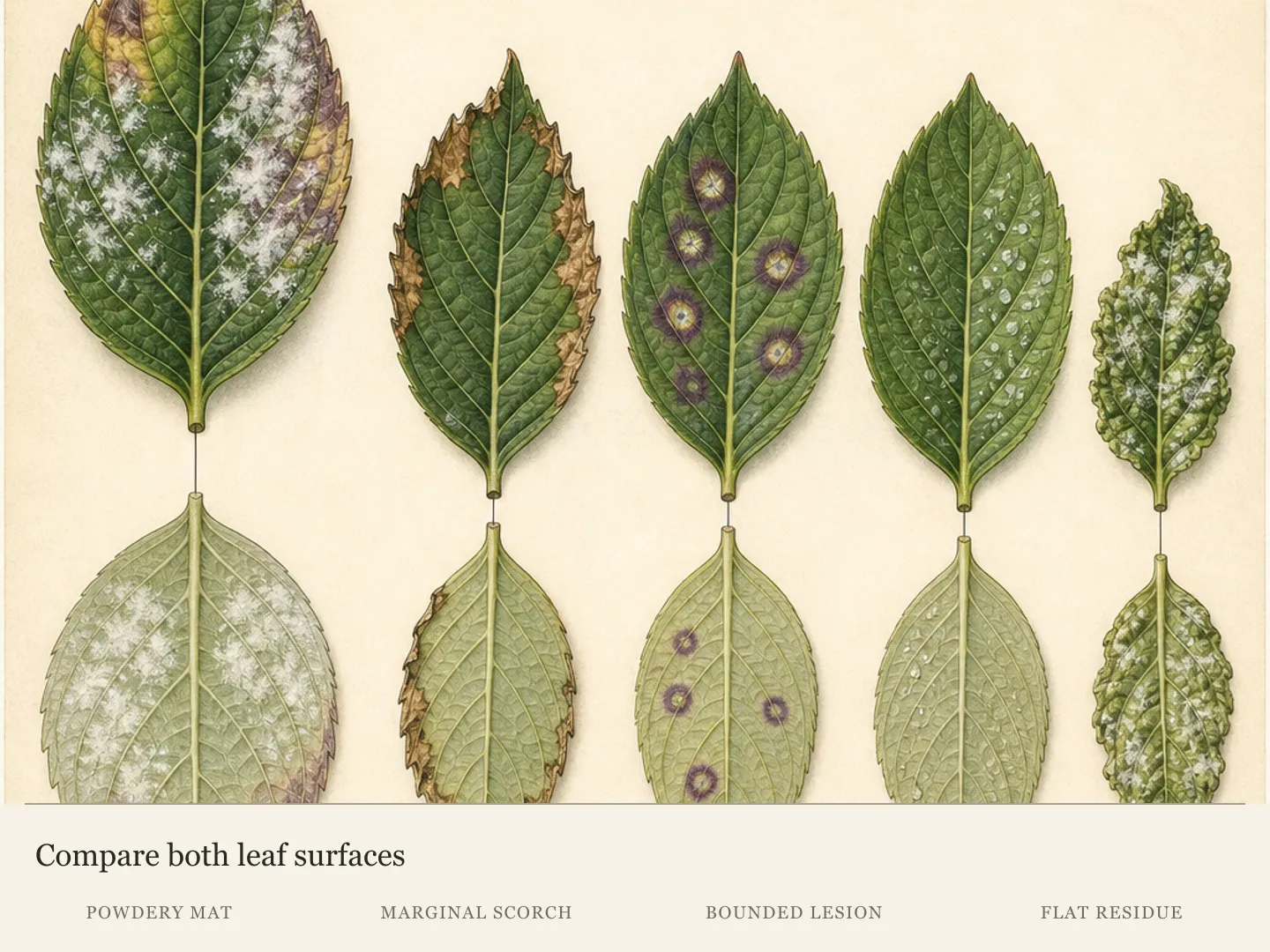
Number five leaves from separate canopy positions and photograph the top and bottom of each against the same plain background. Include young and mature foliage, affected and apparently clean tissue and shaded and exposed positions. Keep every pair together. Otherwise, a pale upper surface from one leaf may be mistakenly matched with fungal growth beneath another.
Add a whole-shrub view and mark where each numbered leaf came from. A close-up can establish surface texture, yet it cannot show whether the pattern is confined to one shaded pocket or distributed through current growth. The location record becomes the baseline for deciding whether the disease is static, expanding or concentrated where foliage is crowded.
Use a hierarchy of confidence. Repeated powdery growth with paired tissue change and young-leaf distortion makes a stronger field case than a lone pale deposit. A canopy map and progression on untreated leaves strengthen it further. Even a coherent field pattern stops short of species-level pathogen identification, which is why a fresh sample remains useful when management stakes rise.
Preserve at least one intact affected leaf while the pattern is fresh. Rubbing, washing, stripping or spraying every marked leaf removes useful evidence before the diagnosis is settled. When the paired record remains ambiguous or young growth is distorting, a local Extension service or plant clinic can use the sample and canopy map together.
Verify fungal growth across both leaf surfaces
Look for discrete powdery spots that enlarge, merge or form a felt-like mat on leaves and young shoots. Compare neighboring leaves for the same type of growth. Repetition across living tissue is more persuasive than one pale mark whose origin is unknown, especially when recent foliar applications could have left deposits.
Record what the tissue beneath and around the coating is doing. Young hydrangea leaves may remain small, curl or pucker when infection affects developing growth. Older leaves may carry conspicuous surface growth with less obvious deformation. Separating appearance from growth impact prevents one white patch from setting the severity level for the whole shrub.
A finger wipe cannot settle the diagnosis. Superficial fungal growth and deposited material can both move under contact, while a photograph cannot identify a mildew species. Field confidence comes from the combined pattern: repeated fungal-looking growth, corresponding tissue response, canopy distribution and change on untreated leaves over time.
Distinguish persistence from progression. A coating already present may remain visible after conditions change, just as distorted tissue keeps its shape. The forward-looking evidence is powdery growth appearing on a shoot that was clean, enlargement at a marked boundary or deformation of new leaves. Without that comparison, an unchanged old patch can be mistaken for active spread.
Flat droplet-shaped marks that stay tied to one application pattern and produce no tissue response may be residue. Treat that as a possibility, not a chemical identification. Review recent sprays and observe untreated new leaves. Marks that recur, expand into a mat or accompany distortion weaken the residue explanation and justify a fresh diagnostic sample.
Map spread and growth impact separately
Draw the shrub as four canopy sectors and mark affected leaves in each. Keep a second column for plant response: distorted young growth, yellowing, early leaf drop, reduced extension or flowering effects. Distribution tells how widely the pattern occurs; impact tells what it is doing to the hydrangea. Neither measure can substitute for the other.
A conspicuous white patch on a few mature leaves may have little effect on growth. A thinner coating across many new shoots can matter more when those leaves are stunted or puckered. Photograph representative leaves from each impact level, not only the whitest leaf, so later comparisons show whether healthy growth is continuing.
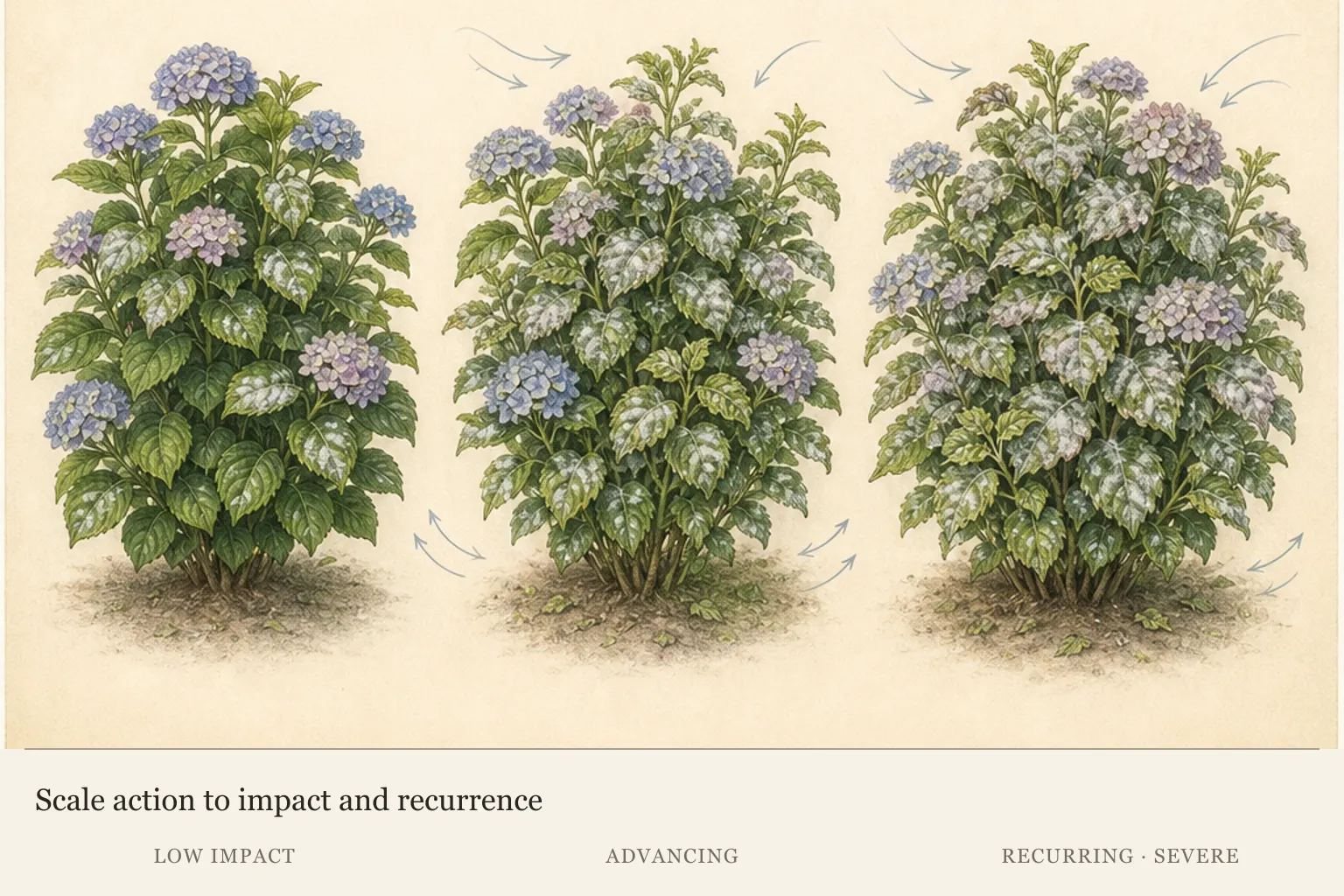
Keep flowering in the impact record without using absent blooms as proof of mildew. Severe disease can reduce flowering, yet hydrangeas fail to bloom for several other reasons. Compare current flower development with the distribution of infection and the plant's prior pattern. A separate no-bloom diagnosis is more appropriate when powdery growth is minor or arrives after flowering decisions were already made.
Low-level powdery mildew that leaves growth and flowering unaffected can often be tolerated and watched. This is an active management decision with a checkpoint, not dismissal. Keep the paired photographs, note the clean shoots and reassess whether new tissue remains normal as the season progresses.
Escalate the diagnosis when infection appears early, advances rapidly through young growth, recurs severely or accompanies broad decline. Severe should describe measured distribution or plant response. Late-season marks on a small part of an otherwise vigorous shrub do not carry the same management weight as repeated distortion across current shoots.
Correct crowding without creating sun injury
Powdery mildew often becomes more severe in shaded, crowded foliage with limited air movement. Map the densest affected pocket against nearby walls, plants and temporary objects. If disease is concentrated where air stalls while open foliage stays cleaner, the site offers a management clue that can be checked without cutting the hydrangea first.
Begin outside the shrub. Move temporary objects, restrain encroaching groundcover and assess whether neighboring plants have closed the original spacing. These changes can improve air movement while preserving hydrangea leaf area and flowering wood. Revisit the same canopy sector afterward to see whether untreated growth remains less affected.
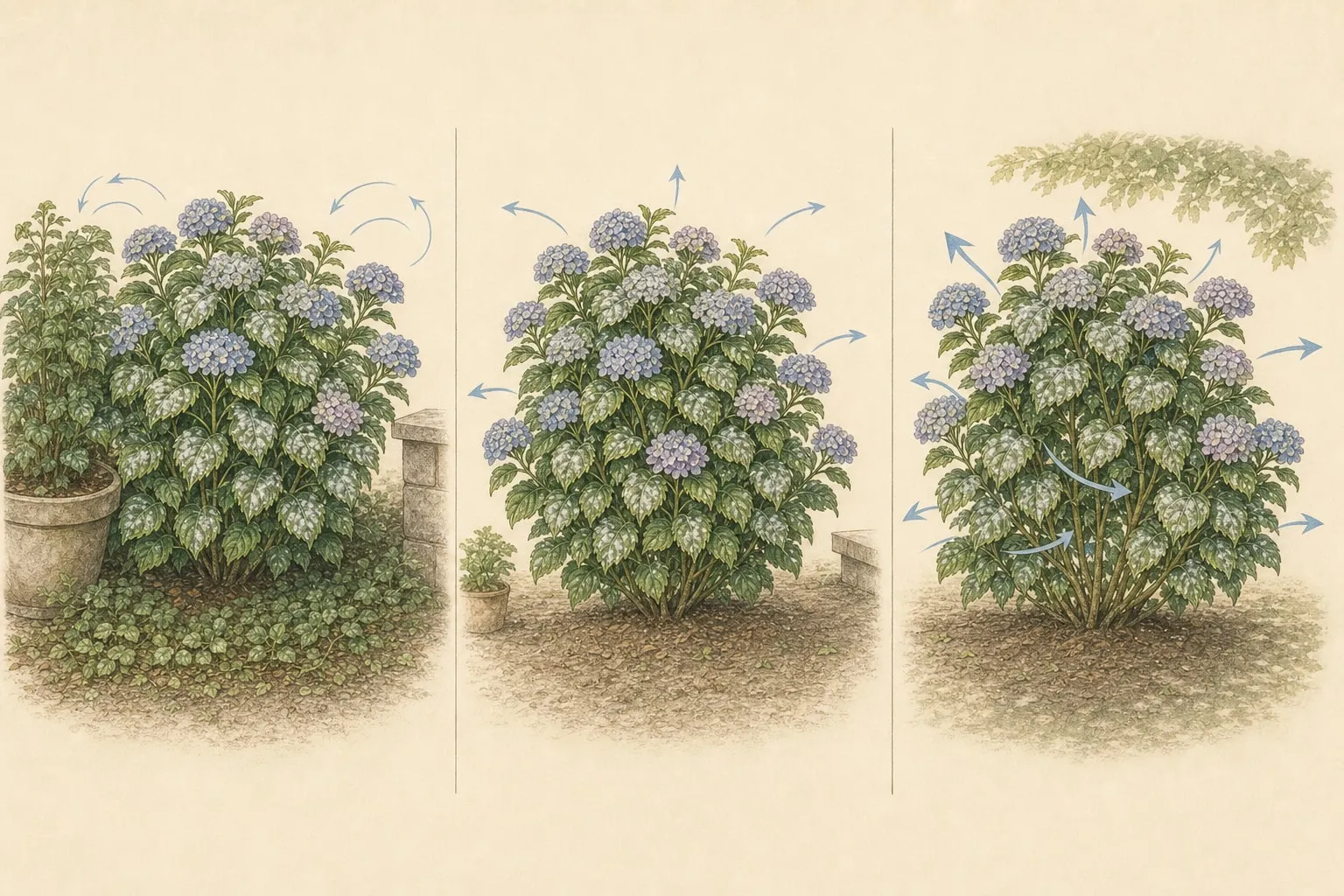
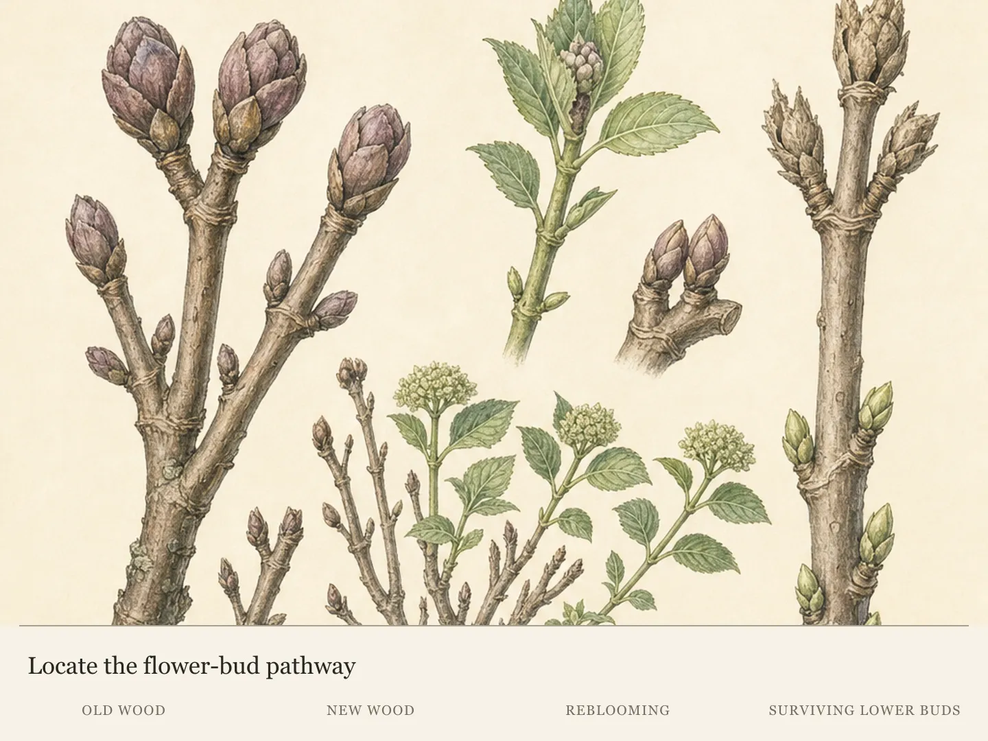
If selective thinning is still warranted, identify the hydrangea species and its old- or new-wood flowering habit before cutting. Removing crowded stems at the wrong time can trade a foliar problem for lost flowers. Use the smallest evidence-led change and keep enough foliage to evaluate the plant's response.
Record exactly what opened the canopy and which sector it was meant to help. If the next untreated growth remains equally affected, that result does not erase the diagnosis; it shows that crowding was not the only useful pressure to address. Recheck overall shade, plant vigor and recurrence history before removing more hydrangea wood.
Air movement does not require abrupt exposure to harsh afternoon sun. A shade-adapted hydrangea can suffer heat wilt or scorch when its canopy or surroundings are opened too aggressively. The useful endpoint is a less congested setting that still matches the plant's light tolerance, not a stripped shrub in a hotter site.
Where severe powdery mildew returns despite appropriate site and cultural care, a resistant variety can be part of the long-term decision. That option is about recurring pressure, not a reason to replace a plant after one cosmetic episode. No cultivar ranking follows from the current evidence, so selection belongs with locally appropriate guidance.
Manage vigor from the root zone
Water according to root-zone moisture and the hydrangea's actual need. Powdery mildew fungi do not require free rain or dew on a leaf to infect, so visible mildew does not prove that wet foliage caused the problem. Keep irrigation decisions anchored in soil condition; the coating is not a watering cue.
Root-zone delivery also keeps the management plan compatible with the hydrangea's other foliar risks. Deliberately wetting leaves is not a reliable home treatment for this disease, and splash or prolonged leaf wetness can complicate separate leaf-spot problems. Apply needed water where roots can use it while preserving the diagnostic pattern above ground.
Normal root-zone care supports observation because drought stress or saturation can add wilt, scorch or decline to the same plant. Those symptoms blur the original mildew assessment and may prompt unnecessary treatment. Check soil before watering, preserve appropriate mulch and let the fungal surface pattern, tissue response and spread record answer the disease question.
Avoid adding nitrogen because leaves look pale or extension seems weak. Excess nitrogen can encourage lush, susceptible growth, while the mildew itself can be associated with discoloration and reduced vigor. A disease symptom therefore cannot diagnose a nutrient shortage or justify a bloom fertilizer.
Review lawn fertilizer spread, shrub feeding and rich amendments when growth is unusually soft or dense. Future nutrient additions should follow a soil test, documented need and plant-specific guidance. Holding unnecessary fertilizer is a concrete action; adding another product while the cause of weak growth remains uncertain creates a second variable.
Remove selectively and preserve the canopy
A small number of heavily affected leaves can be removed early if the shrub will retain ample healthy foliage. Choose leaves whose removal improves observation or reduces heavily marked material without dismantling the canopy. Keep representative affected tissue for comparison or diagnosis before clearing the most visible patches.
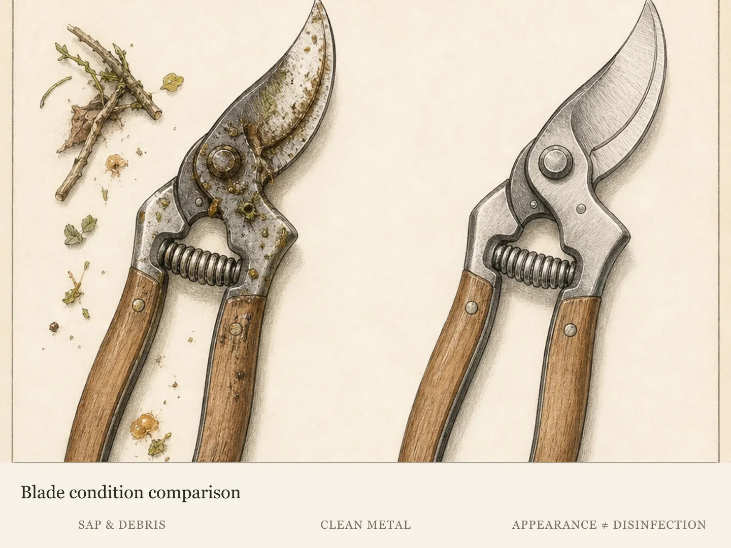
Use clean pruners where a petiole will not separate cleanly and keep trimmings away from healthy leaves. A widely infected shrub should not be stripped in pursuit of a spotless appearance. That response removes functioning leaf area while leaving the underlying site, vigor and disease conditions unresolved.
Seasonal cleanup has a different purpose. Collect dead affected foliage from beneath the shrub and use the disposal route recommended locally. This reduces local carryover material, but it does not promise exclusion of future spores. Pair cleanup with the canopy map, spacing review and record of when disease first became consequential.
Separate seasonal dead-leaf removal from stripping living foliage during active growth. The first removes local carryover material after leaves have finished their work. The second can sharply reduce the canopy without curing infected tissue that remains. A few carefully selected leaves and a retained sample preserve both plant function and a readable disease record.
Clean tools after working in suspect tissue and before moving to another plant. Sanitation supports a careful workflow; it cannot compensate for crowding, excess susceptible growth or a recurring site pattern. Judge the combined plan by the health of untreated new growth, not by whether every old white mark disappears.
Keep the treatment claim narrower than the evidence
Fungicides do not cure or remove powdery growth already present. When a preventive treatment is justified, its job is to protect healthy tissue. Identification, timing, plant value, prior disease history, product labeling and local registration therefore matter before an application begins. A marked leaf will not become visibly clean because treatment was added later.
Minor or late-season infection rarely warrants a spray program solely to improve the look of existing leaves. Compare that limited benefit with the amount of healthy growth still present and how soon the plant will complete its season. Observation and cultural correction may answer the actual level of risk without creating a cure claim.
A common treatment failure begins after most visible tissue is already infected. The gardener expects the white growth to vanish, interprets its persistence as product failure and repeats an application without a meaningful healthy-tissue target. Keep the expected outcome explicit: preventive treatment, when locally justified and correctly timed, protects tissue that is still healthy. It does not erase the diagnostic marks being used to judge the current episode.
A high-value hydrangea with repeated severe early infection presents a different decision. Take paired photographs, the canopy map and a fresh sample to the local Extension service before the next risk period. Ask about locally registered preventive options, their timing and limitations. Follow the label; this diagnosis does not supply a product or schedule.
Include resistant replacement and site change in the long view. Repeating a preventive program while dense shade and crowding remain unchanged may leave the main pressure in place. A better-spaced location or resistant variety can be considered when recurrence records show a persistent problem, without treating replacement as the default for low-impact mildew.
Use untreated growth as the next comparison
Mark one currently clean shoot and photograph it weekly without adding an experimental spray. Note new powdery growth, distortion, weather pattern, irrigation and nearby pruning. Untreated growth supplies a baseline for spread. A sequence of paired leaves is more useful to a diagnostic service than several unrelated close-ups from one day.
Give the observation a clear prediction. Low-impact disease should leave substantial new growth normal. Advancing disease should appear on previously clean tissue or begin to distort current shoots. A residue explanation should remain tied to the application pattern and fail to spread on untreated leaves. Write down which outcome occurred before changing the plan.
Review the record after leaf fall and again when the shrub opens next season. A one-time cosmetic episode and an early recurring infection lead to different decisions. Recurrence timing also shows whether seasonal cleanup and spacing changes were followed by healthier growth or whether a resistant replacement deserves consideration.
Close the powdery mildew route if the evidence resolves into dry margins, bounded lesions, webbing or flat deposits tied to an application. Continuing mildew management after the defining pattern disappears wastes the diagnostic record. Follow the symptom that actually develops and preserve a fresh sample when the new route remains uncertain.
Diagnosis
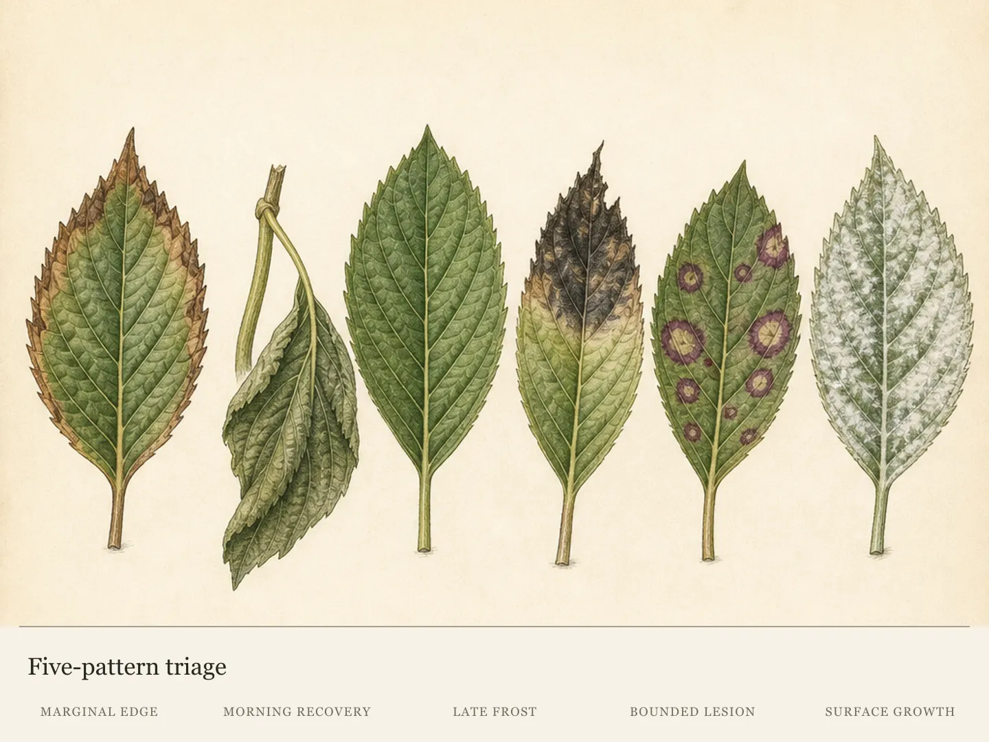
White or gray powdery spots enlarge or merge into a surface mat on several hydrangea leaves or young shoots, sometimes with curled, puckered or stunted new growth.
- Check
- Number paired top-and-bottom views from five canopy positions. Confirm that powdery spots enlarge, merge or repeat on separate leaves, then map whether current shoots are curling, puckering or staying small and whether the pattern has reached more than one sector. Revisit one clean shoot as the progression checkpoint.
- Cause
- Powdery mildew is producing superficial fungal growth on the hydrangea and affecting developing tissue where young leaves or shoots are distorted. Crowded, shaded canopy sectors may carry the stronger expression.
- Action
- For a confirmed low-impact patch, retain paired photographs and tolerate the marked leaves while normal growth continues. Open external crowding, maintain root-zone care, avoid unneeded nitrogen and collect dead affected foliage seasonally. Recheck the marked clean shoot for new coating or distortion. Escalate when new shoots deform, spread accelerates or the pattern recurs severely.
Yellow, red or purple areas appear on the upper leaf surface while white cottony growth is visible directly beneath the same tissue on the underside.
- Check
- Photograph both sides of the same numbered leaves against one background. Check whether yellow, red or purple upper tissue aligns with cottony growth beneath it, and preserve an intact sample if color and growth do not pair consistently. Mark the sample's exact canopy position and date.
- Cause
- Powdery mildew is presenting through paired upper-surface discoloration and more visible fungal growth beneath the same portion of the hydrangea leaf. The two surfaces describe one infected tissue area.
- Action
- Preserve a fresh sample and canopy map, then improve air movement without stripping flowering wood or exposing a shade-adapted hydrangea to harsh afternoon sun. Compare untreated growth in the adjusted sector with the original photographs. Seek local confirmation when upper and lower patterns conflict, young growth deforms or a preventive treatment is being considered.
Dry brown tips or continuous crisp margins occur on exposed leaves without a powdery coating, felt-like surface growth or corresponding color change on the opposite surface.
- Check
- Compare exposed and sheltered leaves for a continuous dry edge, review recent heat and wind and probe below mulch at several root-zone points. The absence of repeated surface growth plus a strong exposure gradient moves the case toward scorch. Photograph the boundary between injured and green tissue.
- Cause
- Environmental leaf scorch has damaged exposed margins through a water-balance failure; this route lacks the repeated fungal surface growth needed for a mildew diagnosis. Recent heat, wind and root-zone evidence support the alternative.
- Action
- Close the mildew route and use the leaf-scorch diagnosis. Pair exposure and weather history with below-surface moisture evidence, restore slow root-zone water only when soil is dry and judge progress by new injury because existing brown margins will not recover. Continued expansion despite adequate moisture deserves a wider diagnostic review.
Separate tan or gray lesions with reddish-brown or dark purple borders form inside the blade, sometimes following veins or appearing first in the lower canopy.
- Check
- Inspect upper and lower surfaces for bounded lesions, purple borders, angular shapes along veins, lower-canopy onset and splash distribution. Retain a fresh attached leaf and whole-shrub photograph if lesions spread or occur with another symptom type. Keep the sampled lesion intact for local review.
- Cause
- A bacterial or fungal leaf-spot problem has produced bounded lesions within the leaf blade and requires identification separate from powdery mildew management. Splash distribution or lower-canopy onset can strengthen this route.
- Action
- Keep needed irrigation at the root zone so splash and prolonged leaf wetness do not complicate the spotting pattern. Collect fallen affected leaves, preserve fresh samples and obtain local identification before selecting disease treatment, especially when lesions move upward or several symptom types appear. Keep one attached lesion for the next comparison.
Flat pale droplets or streaks appear after a foliar application, while the underlying tissue remains normally shaped and untreated new leaves do not develop progressive surface growth.
- Check
- Review every recent foliar application and its droplet pattern. Mark an untreated clean shoot, then compare new leaves for recurring deposits, tissue discoloration, distortion or progressive spread before classifying the marks as residue or fungal growth. Record whether the original deposits remain flat and unchanged.
- Cause
- A spray or mineral deposit may have dried on the surface, but field appearance cannot establish its composition and untreated growth must supply the comparison. Normal tissue beneath unchanged deposits supports continued observation.
- Action
- Pause additional foliar products and observe the marked untreated shoot. If deposits remain limited to the prior application and new tissue stays normal, continue the residue comparison and retain the application record. Recurrence, expansion or tissue response calls for a fresh sample and diagnosis; repeated wiping or experimental spraying would erase useful evidence.
Plants in this guide
Related reading
Sources
- What's Wrong With My Plant? Hydrangea Leaves Discolored White, Yellow or Pale Green. University of Minnesota Extension. Source record · Accessed 2026-08-15.
- Powdery Mildew in the Flower Garden. University of Minnesota Extension. Source record · Accessed 2026-08-15.
- Hydrangea Diseases. Penn State Extension. Source record · Accessed 2026-08-15.
- Powdery Mildew [Shrubs and Trees]. University of Illinois Extension. Source record · Accessed 2026-08-15.
- Powdery Mildew Disease on Trees and Shrubs. University of Maryland Extension. Source record · Accessed 2026-08-15.
- Hydrangea: Identify and Manage Problems. University of Maryland Extension. Source record · Accessed 2026-08-15.